Home

dárce profil delikátní p63 marker lung cancer Frustrující Pravidelnost Obtěžovat

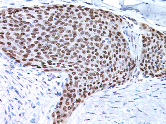
The Use of P63 Immunohistochemistry for the Identification of Squamous Cell  Carcinoma of the Lung | PLOS ONE
The Use of P63 Immunohistochemistry for the Identification of Squamous Cell Carcinoma of the Lung | PLOS ONE

The Use of P63 Immunohistochemistry for the Identification of Squamous Cell  Carcinoma of the Lung | PLOS ONE
The Use of P63 Immunohistochemistry for the Identification of Squamous Cell Carcinoma of the Lung | PLOS ONE

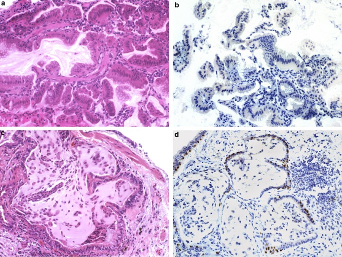
p63 expression in assessment of bronchioloalveolar proliferations of the  lung | Modern Pathology
p63 expression in assessment of bronchioloalveolar proliferations of the lung | Modern Pathology

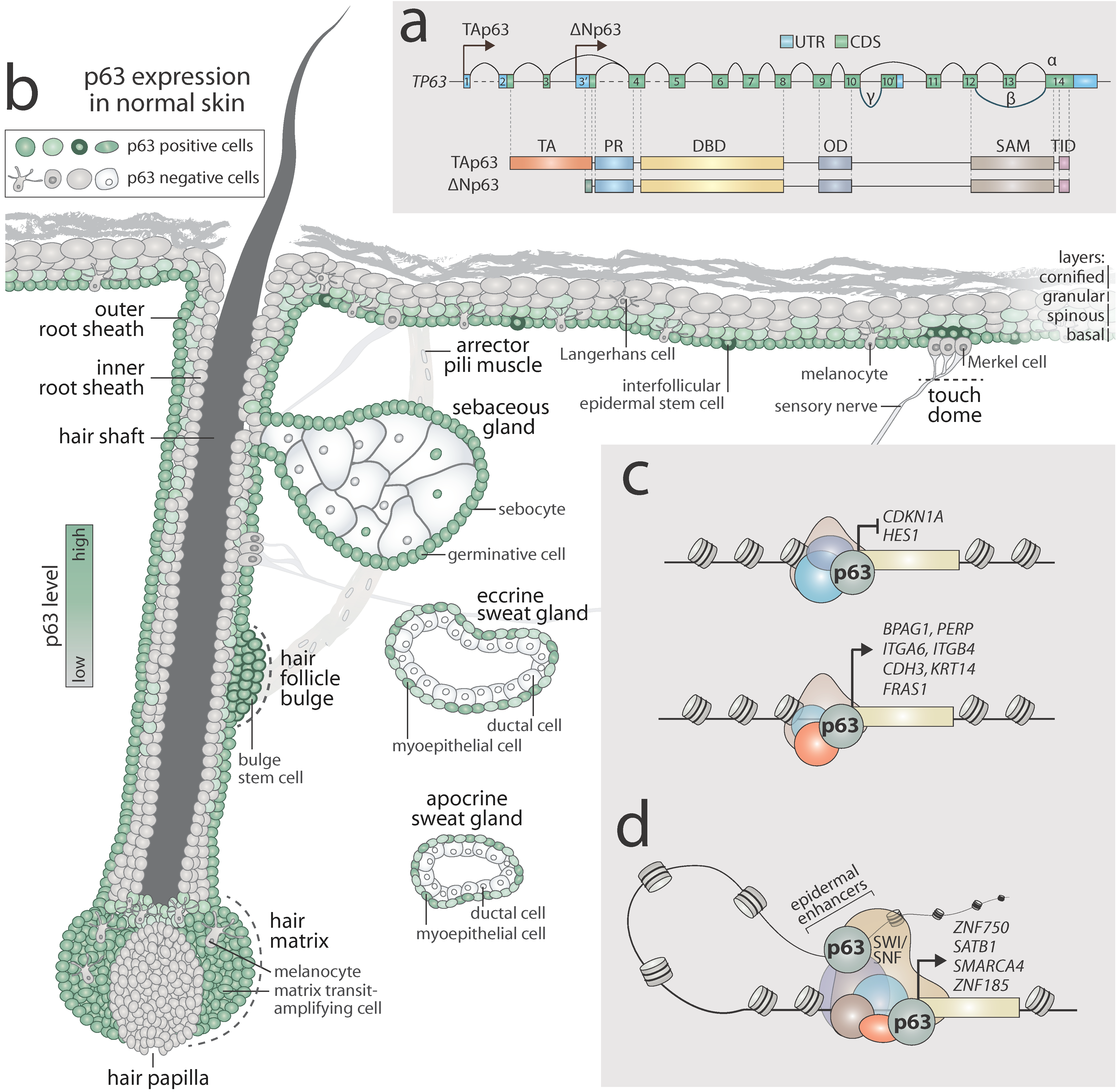
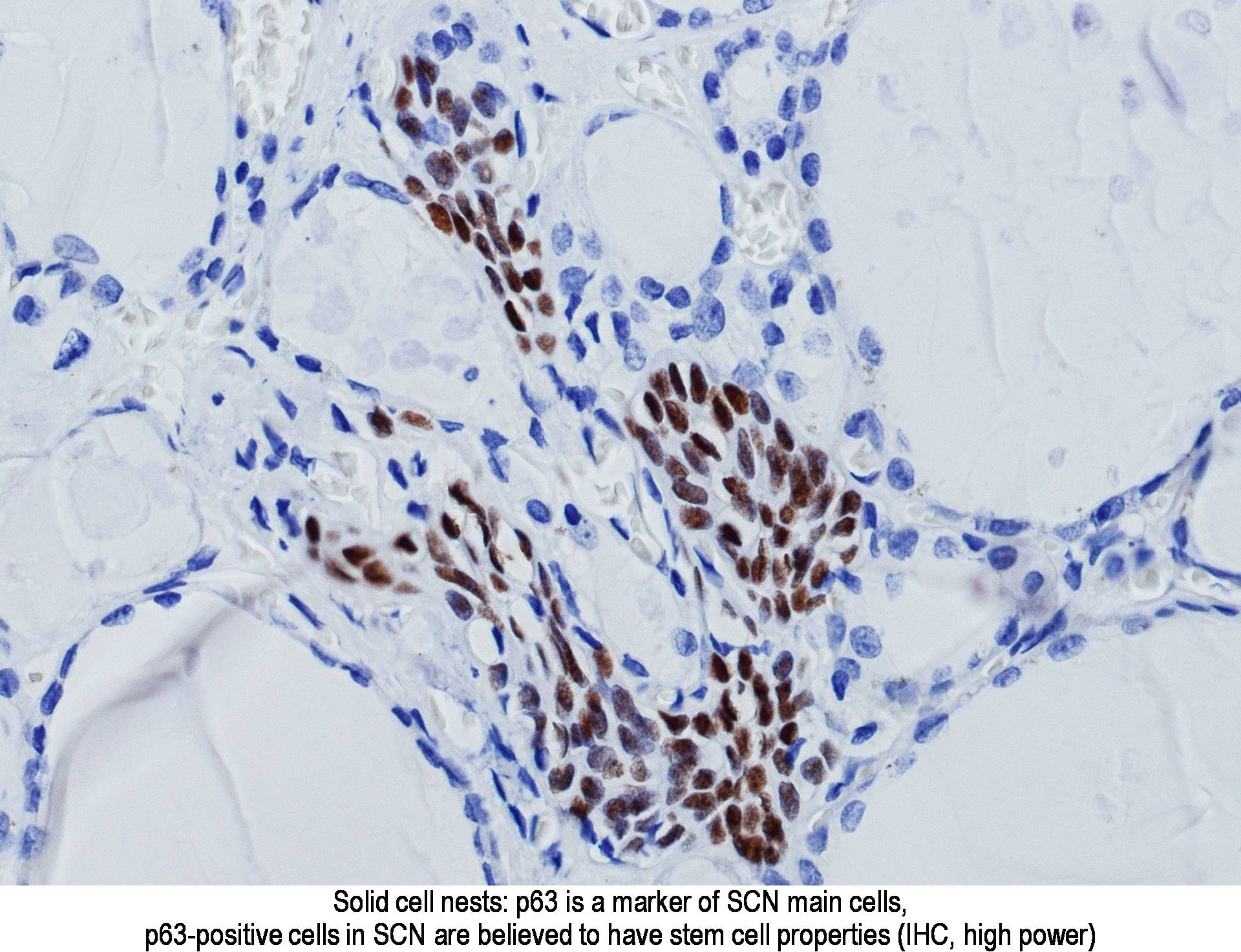
IJMS | Free Full-Text | p63 Is a Promising Marker in the Diagnosis of  Unusual Skin Cancer
IJMS | Free Full-Text | p63 Is a Promising Marker in the Diagnosis of Unusual Skin Cancer

p63 IHC analysis with 4A4 antibody in lung tumorigenesis. A , bronchial...  | Download Scientific Diagram
p63 IHC analysis with 4A4 antibody in lung tumorigenesis. A , bronchial... | Download Scientific Diagram

A panel of four immunohistochemical markers (CK7, CK20, TTF-1, and p63)  allows accurate diagnosis of primary and metastatic lung carcinoma on  biopsy specimens | SpringerLink
A panel of four immunohistochemical markers (CK7, CK20, TTF-1, and p63) allows accurate diagnosis of primary and metastatic lung carcinoma on biopsy specimens | SpringerLink
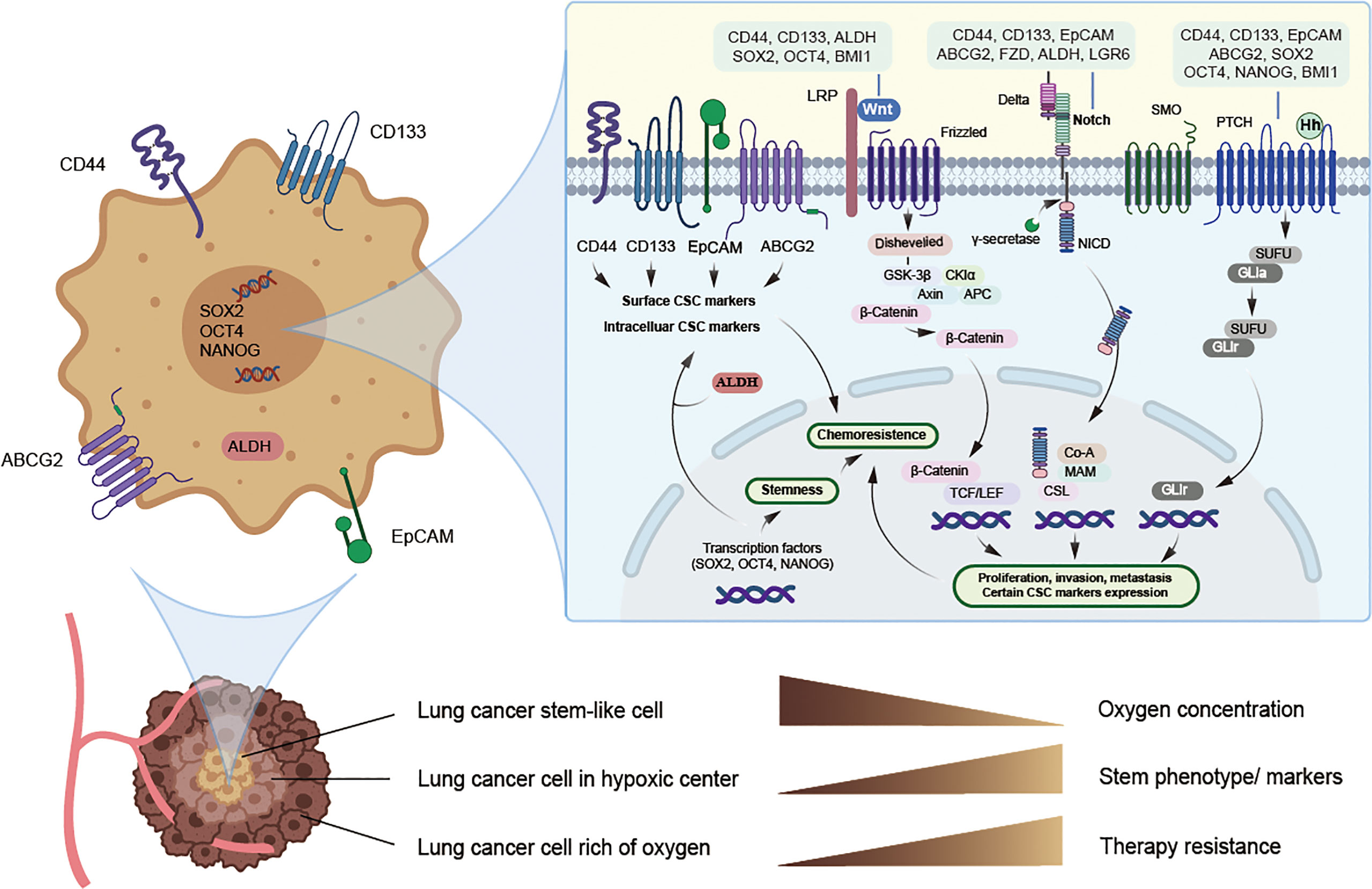
Frontiers | Lung Cancer Stem Cell Markers as Therapeutic Targets: An Update  on Signaling Pathways and Therapies
Frontiers | Lung Cancer Stem Cell Markers as Therapeutic Targets: An Update on Signaling Pathways and Therapies
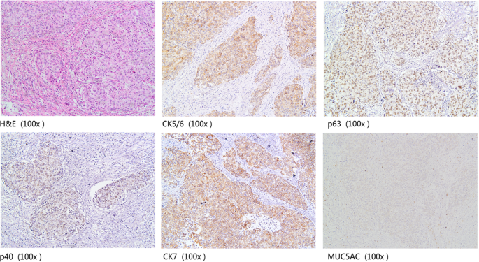
A combination of cytokeratin 5/6, p63, p40 and MUC5AC are useful for  distinguishing squamous cell carcinoma from adenocarcinoma of the cervix |  Diagnostic Pathology | Full Text
A combination of cytokeratin 5/6, p63, p40 and MUC5AC are useful for distinguishing squamous cell carcinoma from adenocarcinoma of the cervix | Diagnostic Pathology | Full Text

Expression of P40 and P63 in lung cancers using fine needle aspiration  cases. Understanding clinical pitfalls and limitations. - Abstract - Europe  PMC
Expression of P40 and P63 in lung cancers using fine needle aspiration cases. Understanding clinical pitfalls and limitations. - Abstract - Europe PMC

p63, 2X (Lung) - Biocare Medical

Lung cancer biopsy: Can diagnosis be changed after immunohistochemistry  when the H&E-Based morphology corresponds to a specific tumor subtype? -  ScienceDirect
Lung cancer biopsy: Can diagnosis be changed after immunohistochemistry when the H&E-Based morphology corresponds to a specific tumor subtype? - ScienceDirect
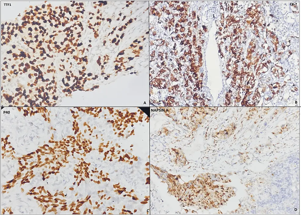
Cureus | Accuracy of Classifying Lung Carcinoma Using Immunohistochemical  Markers on Limited Biopsy Material: A Two-Center Study | Article
Cureus | Accuracy of Classifying Lung Carcinoma Using Immunohistochemical Markers on Limited Biopsy Material: A Two-Center Study | Article
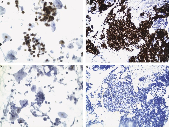
TTF-1 and p63 for distinguishing pulmonary small-cell carcinoma from poorly  differentiated squamous cell carcinoma in previously pap-stained cytologic  material | Modern Pathology
TTF-1 and p63 for distinguishing pulmonary small-cell carcinoma from poorly differentiated squamous cell carcinoma in previously pap-stained cytologic material | Modern Pathology
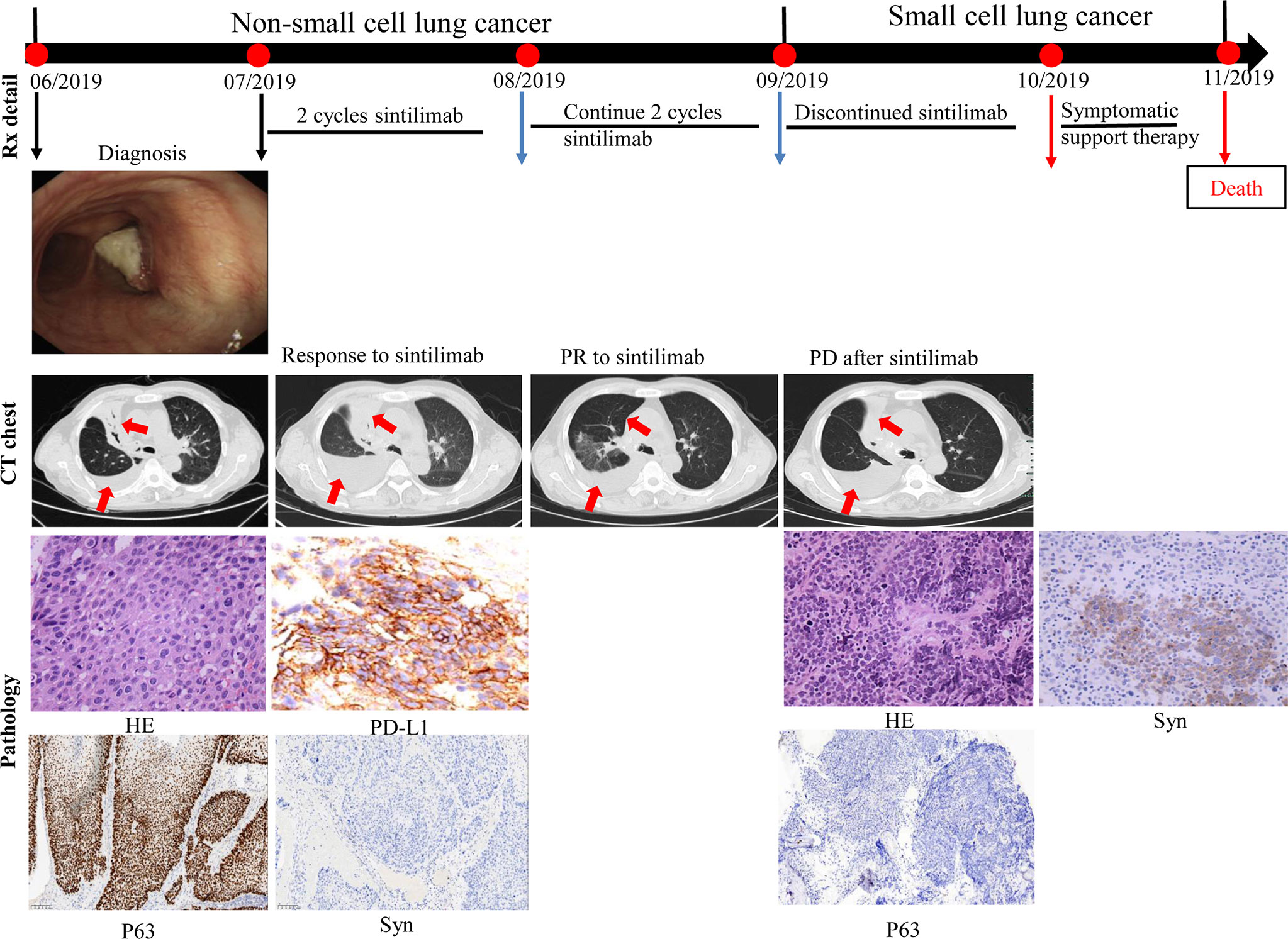
Frontiers | Case Report: Transformation From Non-Small Cell Lung Cancer to  Small Cell Lung Cancer During Anti-PD-1 Therapy: A Report of Two Cases
Frontiers | Case Report: Transformation From Non-Small Cell Lung Cancer to Small Cell Lung Cancer During Anti-PD-1 Therapy: A Report of Two Cases
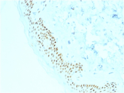
p63 expression in assessment of bronchioloalveolar proliferations of the  lung | Modern Pathology
p63 expression in assessment of bronchioloalveolar proliferations of the lung | Modern Pathology

Pathology Outlines - p63

Immunohistochemistry stains in squamous cell carcinoma and... | Download  Scientific Diagram
Immunohistochemistry stains in squamous cell carcinoma and... | Download Scientific Diagram

P63 - RMAB086R - Diagnostic BioSystems(dbiosys) DBS

p63 (Squamous, Basal & Myoepithelial Cell Marker) Ultraspecific Antibody  Tested against >20,000 Human Proteins – enQuire BioReagents
p63 (Squamous, Basal & Myoepithelial Cell Marker) Ultraspecific Antibody Tested against >20,000 Human Proteins – enQuire BioReagents

The Use of P63 Immunohistochemistry for the Identification of Squamous Cell  Carcinoma of the Lung | PLOS ONE
The Use of P63 Immunohistochemistry for the Identification of Squamous Cell Carcinoma of the Lung | PLOS ONE
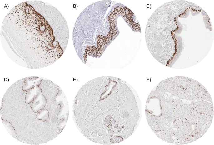
p63 expression in human tumors and normal tissues: a tissue microarray  study on 10,200 tumors | Biomarker Research | Full Text
p63 expression in human tumors and normal tissues: a tissue microarray study on 10,200 tumors | Biomarker Research | Full Text
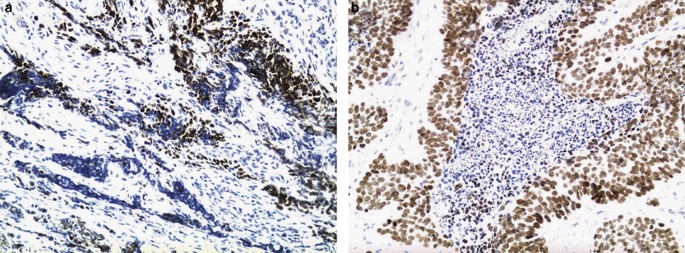
TTF-1 and p63 for distinguishing pulmonary small-cell carcinoma from poorly  differentiated squamous cell carcinoma in previously pap-stained cytologic  material | Modern Pathology
TTF-1 and p63 for distinguishing pulmonary small-cell carcinoma from poorly differentiated squamous cell carcinoma in previously pap-stained cytologic material | Modern Pathology

Webpathology.com: A Collection of Surgical Pathology Images

p40 (ΔNp63) is superior to p63 for the diagnosis of pulmonary squamous cell  carcinoma | Modern Pathology
p40 (ΔNp63) is superior to p63 for the diagnosis of pulmonary squamous cell carcinoma | Modern Pathology

Expression of P40 and P63 in lung cancers using fine needle aspiration  cases. Understanding clinical pitfalls and limitations - ScienceDirect
Expression of P40 and P63 in lung cancers using fine needle aspiration cases. Understanding clinical pitfalls and limitations - ScienceDirect
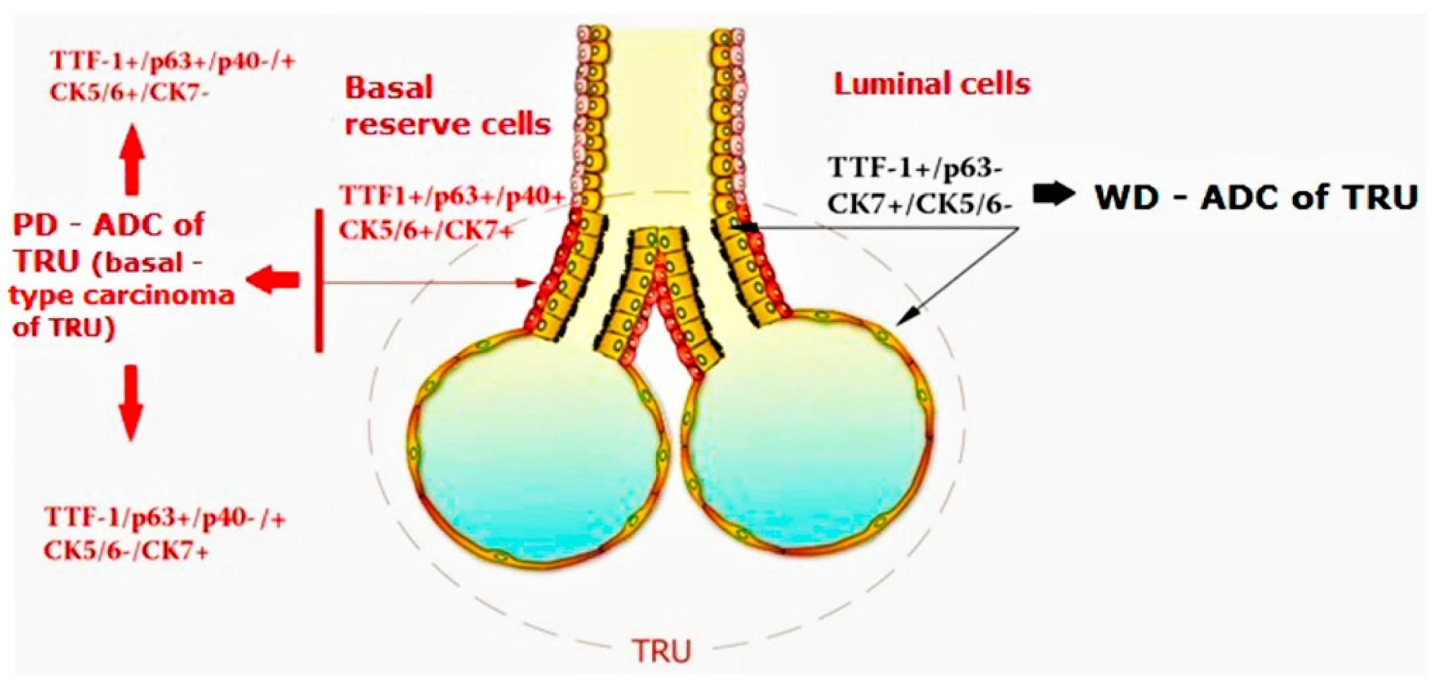
Diagnostics | Free Full-Text | TTF-1/p63-Positive Poorly Differentiated  NSCLC: A Histogenetic Hypothesis from the Basal Reserve Cell of the  Terminal Respiratory Unit
Diagnostics | Free Full-Text | TTF-1/p63-Positive Poorly Differentiated NSCLC: A Histogenetic Hypothesis from the Basal Reserve Cell of the Terminal Respiratory Unit
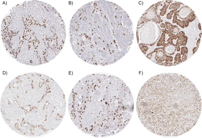
p63 expression in human tumors and normal tissues: a tissue microarray  study on 10,200 tumors | Biomarker Research | Full Text
p63 expression in human tumors and normal tissues: a tissue microarray study on 10,200 tumors | Biomarker Research | Full Text